Home
/ Shoulder Joint Anatomy Diagram - Joints located at the shoulder complex 7 | Download ... - Joints hold the skeleton together and support movement.
Shoulder Joint Anatomy Diagram - Joints located at the shoulder complex 7 | Download ... - Joints hold the skeleton together and support movement.
Shoulder Joint Anatomy Diagram - Joints located at the shoulder complex 7 | Download ... - Joints hold the skeleton together and support movement.. 6 describe briefly the abduction at shoulder joint. It is the major joint connecting the upper limb to the trunk. Clavicle fracture with broken collarbone vector illustration. Shoulder joint of human body anatomy infographic diagram with all parts including bones ligaments muscles bursa cavity capsule cartilage membrane for medical science education and health care. There are two ways to categorize joints.
The first is by joint function, also referred to as range of motion. The multiple ligaments and tendons around the shoulder must be strong to bind the shoulder joints together and encapsulate them in a tough but flexible structure. Clavicle fracture with broken collarbone vector illustration. Corey chakarun from shin imaging in california. Shoulder joint is the most mobile joint of the human body.
Frozen Shoulder: you'd rather it was just cold from www.sghs.org Assessment | biopsychology | comparative | cognitive | developmental | language | individual differences | personality | philosophy | social | methods | statistics | clinical | educational | industrial | professional items | world psychology |. Dislocation of the shoulder is extremely painful and may require surgical repair or even cause permanent damage. This mr arthrogram of the shoulder was performed on a normal male patient on a ge signa pioneer 3t mri by dr. Diagram of the different insertions of the anterior capsule as seen on the axial plane (arrowheads). Medical and anatomical labeled scheme with clavicle fracture, acromion, humeral head, scapula and humerus. The glenohumearal joint has a greater range of motion than any other joint in the body. The shoulder is actually composed of four joints, namely glenohumeral joint, acromioclavicular joint, sternoclavicular joint and scapulothoracic joint. The shoulder joint is the connection between the chest and the upper extremity.
It is the major joint connecting the upper limb to the trunk. Human anatomy for muscle, reproductive, and skeleton. There are two ways to categorize joints. The shoulder joint is vulnerable to dislocations from sudden jerks of the arm, especially in children before strong muscles have developed. Webmd's shoulder anatomy page provides an image of the parts of the shoulder and describes its function, shoulder problems, and more. Corey chakarun from shin imaging in california. Related posts of muscle anatomy of shoulder joint. In human anatomy, the shoulder joint comprises the part of the body where the humerus attaches to the scapula.1 there are two kinds of cartilage in the joint. In common usage, shoulder joint mostly refers to the glenohumeral joint, the major joint of the shoulder but can also include acromioclavicular joint. Clavicle fracture with broken collarbone vector illustration. Diagram of the different insertions of the anterior capsule as seen on the axial plane (arrowheads). Start studying shoulder joint anatomy. Related online courses on physioplus.
Shoulder joint is the most mobile joint of the human body. Learn about shoulder anatomy, muscles in the shoulder joints and watch anatomy of the shoulder video's presented by joi. Describe the structure of the shoulder should begin with bone parts that include: Click now and learn everything about its anatomy and function at kenhub! Editor · aug 6, 2017 ·.
File:Shoulder joint back-en.svg - Wikimedia Commons from upload.wikimedia.org This acts as the bony framework by which the muscles of the chest, upper back and shoulder connect the upper limb to the trunk of the body and control it's movements.the clavicle connects to the sternum via the sternoclavicular joint and to the scapula by. The shoulder is actually composed of four joints, namely glenohumeral joint, acromioclavicular joint, sternoclavicular joint and scapulothoracic joint. Normal anatomy, variants and checklist. 7 draw labelled diagram showing the relations of shoulder joint. The human shoulder is the most mobile joint in the body. Clavicle fracture with broken collarbone vector illustration. There are two ways to categorize joints. Visualization of the humeral head and joint space free of superimposition.
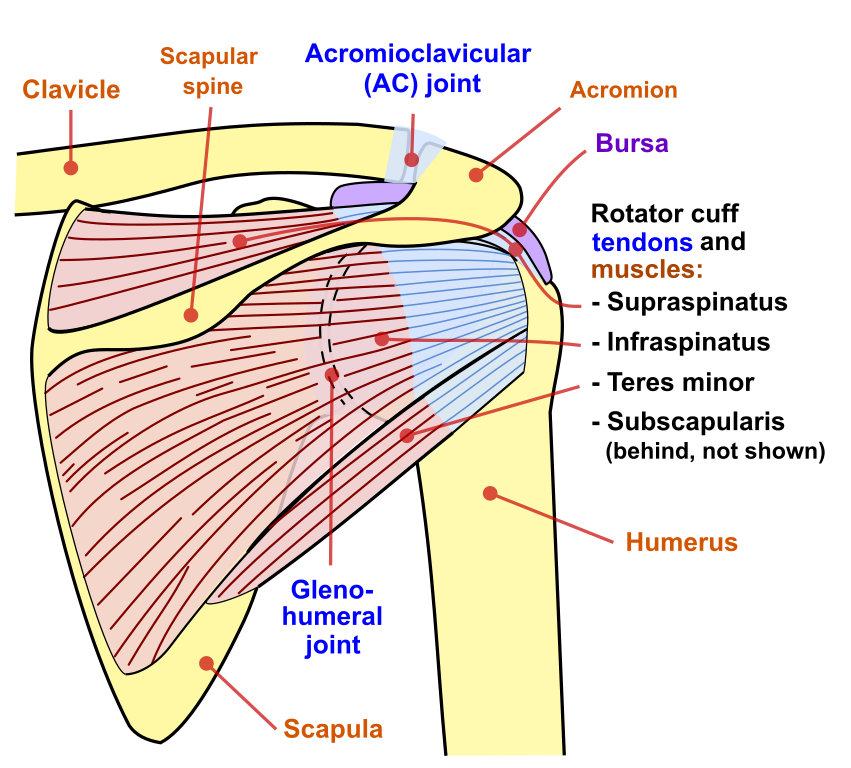
• during abduction of the shoulder joint, the supraspinatus tendon is exposed to friction against the acromion.
The multiple ligaments and tendons around the shoulder must be strong to bind the shoulder joints together and encapsulate them in a tough but flexible structure. The human shoulder is the most mobile joint in the body. Related posts of muscle anatomy of shoulder joint. Shoulder joint of human body anatomy infographic diagram with all parts including bones ligaments muscles bursa cavity capsule cartilage membrane for medical science education and health care. This diagram here just shows the joint capsule itself. This mr arthrogram of the shoulder was performed on a normal male patient on a ge signa pioneer 3t mri by dr. The shoulder joint is vulnerable to dislocations from sudden jerks of the arm, especially in children before strong muscles have developed. Just remember the articulating surfaces. Labeled human shoulder bone anatomical vector illustration diagram poster. This diagram with labels depicts and explains the details of anatomy of the shoulder joint. Equally extensive are the muscles affecting the shoulder movement, including: You can see it enclosing the glenohumeral joint and you can see its attachment on the anatomical neck that's the shoulder joint. There are two ways to categorize joints.
The human shoulder is the most mobile joint in the body. Medical and anatomical labeled scheme with clavicle fracture, acromion, humeral head, scapula and humerus. This attaches to the upper and posterior end of the clavicle and cartilage of the 1st rib purpose: Visualization of the humeral head and joint space free of superimposition. In human anatomy, the shoulder joint comprises the part of the body where the humerus attaches to the scapula.1 there are two kinds of cartilage in the joint.
Shoulder joint; Glenohumeral Joint from classconnection.s3.amazonaws.com Three bones come together at the shoulder joint. Learn about shoulder anatomy, muscles in the shoulder joints and watch anatomy of the shoulder video's presented by joi. 8 name the arteries and the. Human anatomy for muscle, reproductive, and skeleton. The shoulder joint is vulnerable to dislocations from sudden jerks of the arm, especially in children before strong muscles have developed. Webmd's shoulder anatomy page provides an image of the parts of the shoulder and describes its function, shoulder problems, and more. It is the major joint connecting the upper limb to the trunk. In common usage, shoulder joint mostly refers to the glenohumeral joint, the major joint of the shoulder but can also include acromioclavicular joint.
8 name the arteries and the.
The shoulder joint is the connection between the chest and the upper extremity. This diagram with labels depicts and explains the details of anatomy of the shoulder joint. Joints hold the skeleton together and support movement. This acts as the bony framework by which the muscles of the chest, upper back and shoulder connect the upper limb to the trunk of the body and control it's movements.the clavicle connects to the sternum via the sternoclavicular joint and to the scapula by. Editor · aug 6, 2017 ·. Three bones come together at the shoulder joint. In this article, we shall look at the anatomy of the shoulder joint and its important clinical correlations. Equally extensive are the muscles affecting the shoulder movement, including: The shoulder joint is vulnerable to dislocations from sudden jerks of the arm, especially in children before strong muscles have developed. Shoulder anatomy is a remarkable combination of strong bones, flexible ligaments and tendons, and reinforcing cartilage and muscles. Webmd's shoulder anatomy page provides an image of the parts of the shoulder and describes its function, shoulder problems, and more. Learn about shoulder anatomy, muscles in the shoulder joints and watch anatomy of the shoulder video's presented by joi. The shoulder joint is formed where the humerus (upper arm bone) fits into the scapula (shoulder blade), like a ball and socket.
Shoulder joint of human body anatomy infographic diagram with all parts including bones ligaments muscles bursa cavity capsule cartilage membrane for medical science education and health care shoulder anatomy diagram. Shoulder joint of human body anatomy infographic diagram with all parts including bones ligaments muscles bursa cavity capsule cartilage membrane for medical science education and health care.

